

Cancer Cell Panel & In Vitro Oncology Assays
Overview
We offer a diverse in vitro screening panel of over 600 authenticated, well-validated human cancer cell lines across 30 cancer types. Our cancer cell line panel provides powerful tools for cancer research and drug discovery. Key features of the WuXi cancer cell panel include:
- High quality cell banking system: mycoplasma-proof and STR verified
- Single agent and combination compound screening for potency (% inhibition & IC50) and synergy (combination index CI)
- Professional design with stringent quality control of study results
- A database of cell growth curve and Standard of Care (SoC) validation data provides guidance for cell model selection
- 9 drug-resistant cell lines
- Over 150 engineered cell lines
- 95 PDX-derived cell lines
- More than 100 mouse lines, covering 20+ cancer types
Large Scale Human Cancer Cell Lines for Cell Panel Services
A wide range of 600+ human cancer cell lines is available for oncology drug discovery programs. The cell banking system is a two-tiered banking system with mycoplasma testing and STR validation. Established cell growth data and standard of care profiling enhance the quality of study. The table shows the distribution of these human cancer cell lines, sorted by the origins of derived organs/tissues.
Cancer Cell Line | Number of Cell Lines | % of Total |
Lung | 120 | 31.7 |
Breast | 66 | 17.5 |
Hematopoietic/lymphoid tissue | 44 | 11.6 |
Large intestine | 35 | 9.3 |
Liver | 15 | 4.0 |
Skin | 11 | 2.9 |
Connective/soft tissue | 11 | 2.9 |
Stomach | 9 | 2.4 |
Pancreas | 9 | 2.4 |
Brain | 9 | 2.4 |
Kidney | 8 | 2.1 |
Bone | 7 | 1.8 |
Upper aerodigestive tract | 7 | 1.8 |
Bladder | 6 | 1.6 |
Ovary | 6 | 1.6 |
Prostate | 5 | 1.3 |
Thyroid | 3 | 0.8 |
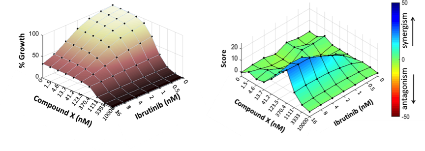
Synergistic effects of Compound X with Ibrutinib in TMD8
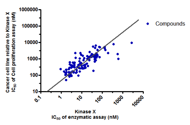
Potency Correlation of Compounds (kinase assay & cell proliferation assay)
Cell line-derived xenograft (CDX) tumor models
Overview
- A large collection of cell line-derived xenograft (CDX) tumor models
-Over 320 validated models, SOC data available - World leading imaging based orthotopic/metastatic CDX platform
-28 models covering 16 tumor types
Cell Line-Derived Xenograft (CDX) Tumor Models: 320
Tumor Types | Cell lines |
Bladder | T24/83, RT112/84, UM-UC-3, J82 |
Brain | U87*, LN-229* |
Breast | Cal51*, BT-474*, HCC1954*, MDA-MB-231*, MDA-MB-468*, HCC1806*, MCF7*, HCC1937, MDA-MB-436*, SUM-52Pe, JIMT-1*, MDA-MB-361*, HCC-70, SUM 159PT, MFM-223, T47D*, HCC1428, Z-75-1* |
Cervical | SW756 |
Colorectal | Colo205*, HT29, HCT-116*, HCT-15*, LS180*, LS174T*, SW620*, HT55*, DLD-1, Lovo, COLO-320DM, RKO, Caco-2, T84, WiDr, SW480, SW 48, LS411N, COLO 201 |
Duodenal | HuTu80 |
Esophageal | T.Tn, OE21* |
Gastric | NUGC4*, SNU-1, SNU-16*, MCG-803, NCI-N87*, SNU-5, MKN45*, HS 746T |
Head & Neck | Cal 27, RPMI2650, SCC-9, Fadu* |
Leukemia | HEL 92.1.7*, MV4-11*, CCRF-CEM, HL-60, K562, KG-1, MOLT-4, Kasumi-1*, Nalm-6*, THP-1, SET-2, PL-21, MLK-1 |
Lymphoma | Daudi*, SU-DHL-2*, SU-DHL-10, Granta-519, Maver-1, Mino, NAMALWA, Raji, Ramos, RL, WSU-DLCL2, Z-138, U379, DoHH2, Farage*, Pfeiffer, REC-1*, OCI-LY10, TMD-8* |
Tumor Types | Cell lines |
Liver | BEL-7404*, Hep3B*, Huh 7*, HepG2, SNU-182, QGY-7703, SNU-398, JHH-7*, MHCC97H* |
Lung | NCHI-H1650, NCI-H226, NCI-H460, PC9*, A549*, Calu-1, Calu-6*, EBC-1, A427, NCI-H647, MSTO-211H, NCI-H292, NCI-H322, NCI-H358*, NCI-H441, NCI-H520, NCI-H1957*, NCI-H2228, SK-MES-1, NCI-H1229, HCC-827*, HCC400646, NCI-H1703, NCI-H727, NCI-H2122*, DV-90, NCI-H69*, NCI-H1417*, NCI-H446, NCI-H526*, NCI-H82, SHP-77, Calu-3, NCI-H2170 |
Melanoma | A375*, SK-MEL-24, SK-MEL-28, COLO 829*, SK-MEL-5, MDA-MD-435S |
Myeloma | KMS-28BM, KMS-11, MOLP-8*, NCI-H929, RPMI 8226*, MM.1S* |
Neuroblastoma | CHP-134*, SK-N-AS*, BE(2)-C*, SH-SY5Y |
Ovary | A2780*, SK-OV-3*, ES-2*, OVCAR-3, TOV21G |
Pancreas | BxPC3*, Mia-pa-ca-2*, KP4, PANC-1*, CAPAN-1, Panc 02.13* |
Prostate | PC-3*, DU145, LNCap*, VCaP, 22RV.1 |
Renal | 786-O* ACHN, Caki-1, SK-NEP-1, A498 |
Sarcoma | HT1080, A673, KHOS/NP, 143B, SJSA-1*, SAOS-2, A204, G401 |
Skin | A431* |
*: validated with SOC compound
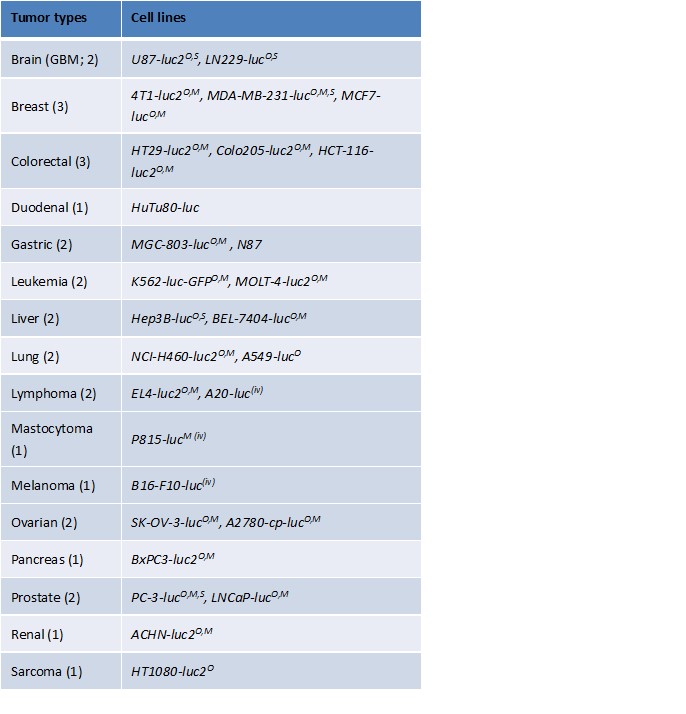
Luciferase Labelled Orthotopic/Metastatic Tumor Models
- Covering 16 tumor types
- Bioluminescent imaging system in SPF barrier
Immuno-oncology
One stop service to enable cancer immunotherapy
Outstanding Platform to Enable Cancer Immunotherapy with One Stop Service
- Immunological function assessment
- Tumor microenvironment analysis
- In vivo efficacy evaluation on animal models
- NGS to understand TCR/BCR Repertoire
- Patient TILs analysis
Immuno-oncology In Vitro Functional Assays
- Immune cell activation assays
– T/B/NK cell activation - Macrophage differentiation assays
– M1/M2 macrophage differentiation
– DC differentiation
– MDSC induction - T cell polarization assays
– Treg/Th1/Th2/Th17 differentiation - T cell cytotoxicity assay
– Cytotoxic T lymphocyte assay (OT-1 system) - Mixed Lymphocyte Reaction
- Antibody-Drug Conjugate (ADC) binding
- Immune cell suppressive assay
– MDSC/Treg/Tumor-T suppressive assay - Antibody mediated immunological assay
– ADCC/CDC/ADCP
Assay | WuXi capibility (Human/Mouse) | |
Call activation | T cell | Yes (H/M) |
B cell | Yes (H) | |
NK cell | Yes (H) | |
Cell differentiation/ polarization | M1/M2 macrophage | Yes (H/M) |
MDSC | Yes (H) | |
DC | Yes (H/M) | |
Treg, Th1, Th2, Th17 polarization | Yes (H) | |
Mixed Lymphocyte Reaction | DC co-culture with T | Yes (H/M) |
Suppressive Assay | MDSC suppressive assay | Yes (H) |
Treg suppressive assay | Yes (H) | |
Tumor cell+immune cell co-culture | Yes (H) | |
Cell cytoxicity | Cytotoxic T lymphocyte assay (OT-1 system) | Yes (M) |
NK cytotoxicity | Yes (H) | |
ADCC, ADCP | Yes (H) | |
CDC | Yes (H) | |
Cell exhaustion | T cell exhaustion | Yes (M) |
Immuno-oncology In Vitro Technical Competence
Immune primary cell profiling in tumor infiltrating leukocyte (TIL) and other tissues (spleen, blood, lymph node, liver) profiling: (1) Immune cell population analysis (T, B, NK, DC, Myeloid, Macrophage, Granulocyte, CD4+T, CD8+T, Treg, Th1/2/17, naïve/memory/effector T, g/mMDSC, M1/M2, etc.) (2) Functional and intracellular marker analysis (PD-1, PD-L1/2, CTLA-4, TIM-3, LAG-3, OX40, 4-1BB, ICOS, Ki-67, IL-2, IL-17, IFNγ, TNFα, Granzyme B)
Biomarker
- Soluble biomarker – ELISA, ELISPOT
- Cell-based biomarker – FACS
- Tissue-based biomarker – Multiplex IHC
- Gene expression profiling – RNAseq, NanoString
Tumor Infiltrating Lymphocytes (TILs) Analysis on Human and Murine Tumor Tissues
- CD4, CD8, MDSC, M1/M2, DC cell, NK cell, B cell
- PD1, TIM3, LAG3, OX40, 4-1BB
- Treg, Th1,2,3, and17
In Vivo Efficacy Evaluation on Animal Models Syngeneic Murine Models
- Immune-checkpoint humanized models
- Immune-Avatar humanized models PBMC humanized models
- HSC humanized models
TM (Tumor Microenvironment) Analysis for Immunoscore by FACS, IHC and IF
CTLs, NK, Tregs, MDSC, and TAM
Lymphoid Tissue and Peripheral Blood Analysis
- Immune cell populations and subsets – frequency, activation, and differentiation
- Cytokine and chemokine analysis
- Anatomical localization ofin vivo anti-tumor immune responses
